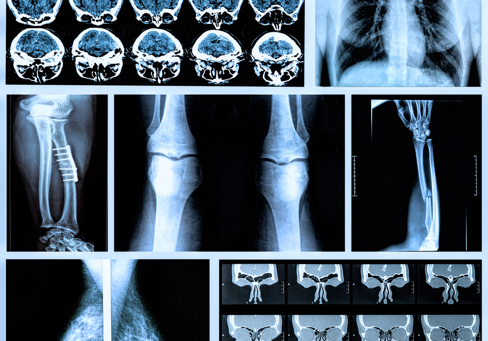
X-ray
Some Areas Where X-rays are Used:
RADIOSCOPY: It is examined by an organ or cismin X rays with the help of a fluorescent screen. Radioscopy is the examination of the shadows brought by the X rays on a fluorescent screen made of barium platinocyanurium or tungsten. Radioscopy provides a quick examination of the whole body, in the form of every posture and examination of organs from all angles.
RADIOGRAPHY: X-rays on a sensitive film in a box that passes X-rays only, leaving a mark and taking advantage of this feature. (The box used for this work is made of a light mineral such as aluminum).
The X-ray film used for the radiography is usually placed between two plates that become fluorinated by the action of X-rays. These plates greatly enhance the effect of X-rays and shorten the exposure time. The radiograph gives the images of the chest and heart without the need for special preparation thanks to the contrast provided by the air in the lung air fins. The calcium-loaded skeleton is well documented in the radiography, with abnormal formations (kidney and gall stones, calcified lymph nodes, etc.) with too much calcium salt in it.
RADIOMETHALOGRAPHY: Radiography is used to examine the composition or structure of metal parts without disturbing.
It is based on the same principles of physics as medical radiography. The ability to vary the chemical composition and to differentiate the X-rays of various parts of a metal part in order to inculcate defects in the inner structure of the mine is exploited. In particular, it absorbs less X-rays and allows the determination of voids and less dense parts on the film that appear darker than normal regions. Foreign materials which are also mixed in the same part and which are different from the minerals in which the number of absorption layers are made are also seen as lighter or darker stains on the film. Radiometry also makes it easier to check whether some of the copper alloys or mines (such as high-sorption lead) are homogeneous in structural and chemical maintenance.
TOMOGRAPHY: It is the procedure to take an organ and an organism section with x-ray filmini. In fact, a slice of thin film is about 1-2 cm in thickness. Thus, a particular organ, for example lung, can be examined in horizontal or horizontal and vertical longitudinal planes.
To make the tomography, the X-ray-producing tape and the sensitive film are subjected to various displacement movements so that only the shapes on a certain plane that coincides with the axis of this displacement movement appear in the film; in front of, behind, on, under, and so on. The shapes found are not clearly visible. In other words, the sensitive film is hardly affected at all, but it appears in very faint lines.